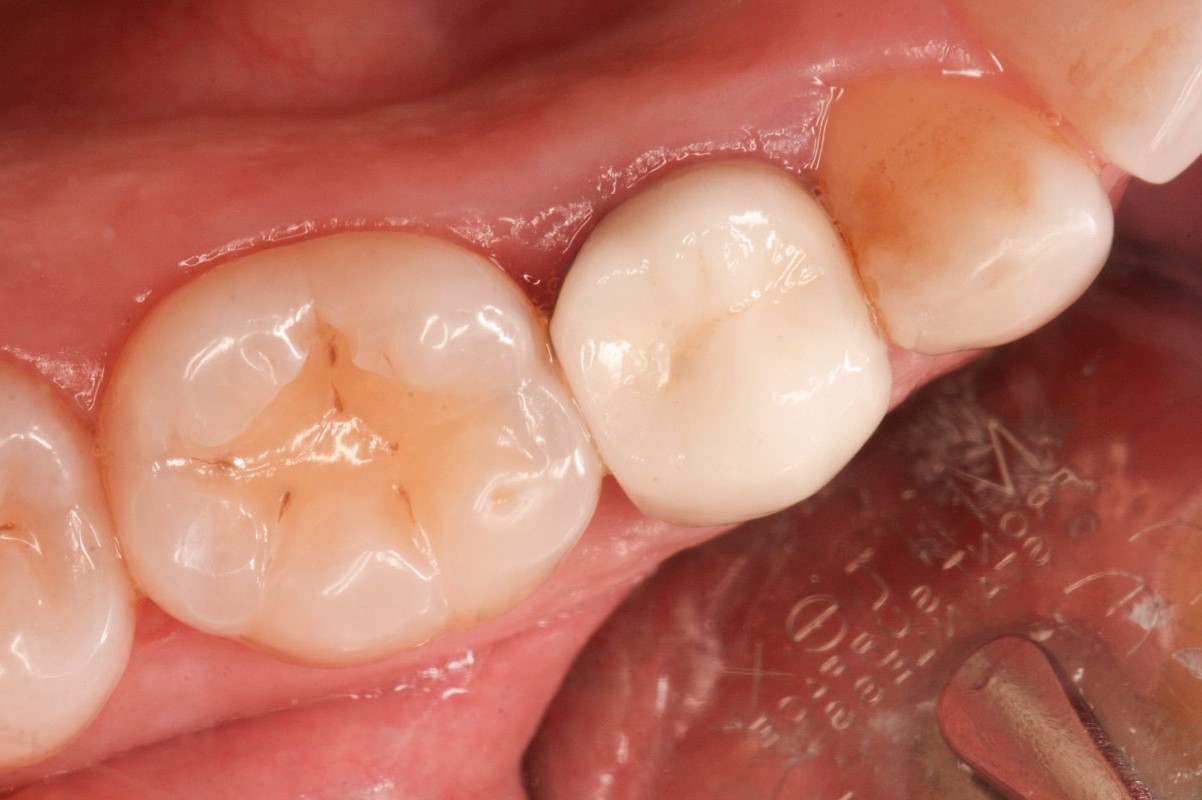
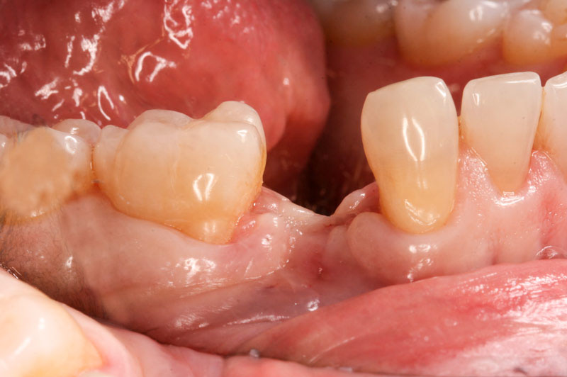
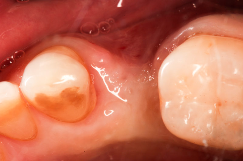
This young man had an ugly looking crown over a dead tooth which could not be saved.
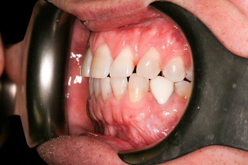
We extracted the tooth and placed a small graft to preserve whatever bone we had. This is what it looked like 6 months later.
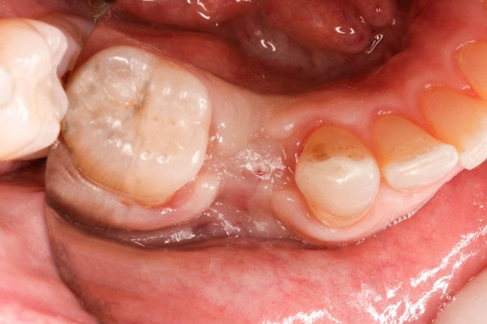
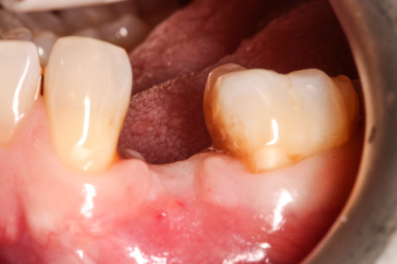
We had a surgical stent made, and a 3-D bone scan was ordered to position the implant exactly. We used the same stent to prepare the bone for the implant.

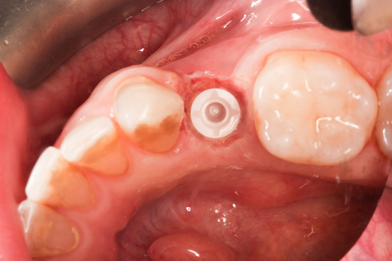
Due to lack of bone anatomy, we separated the gums to visualize the placement and to confirm the position of the implant.
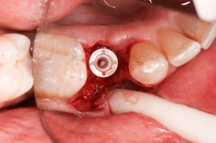
Considering the lack of bone, the implant was placed slightly deeper and the area was covered by the gums and closed up with sutures. (This is not normal practice with proper bone. The implant usually stays exposed at the level of the gum.)
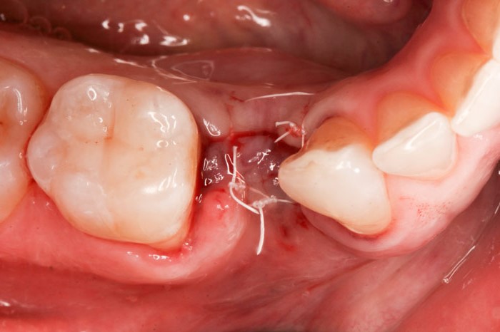
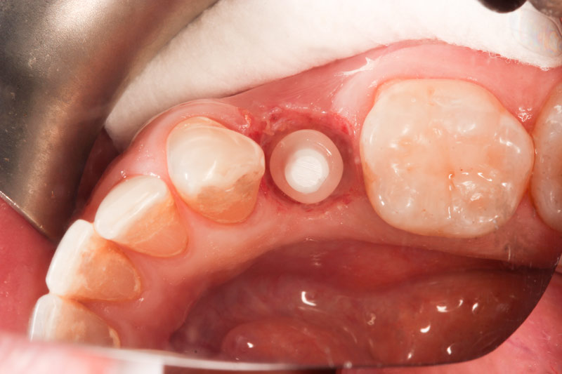
Five months later the area has healed and the implant has integrated with bone.
The waterlase dental laser was used to expose the implant non-surgically. Due to the excellent biocompatibility of zirconia, the gums are not inflamed at all.
The waterlase is also used to clean and debride the top part of the implant which will be receiving the cemented abutment (the post upon which the actual crown will be placed). This procedure makes the implant look as shiny as the moment it came out of the packaging.

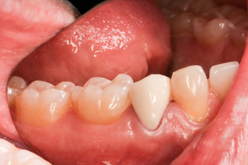
The post is cemented.
The post may still be slightly modified before the impression is taken.

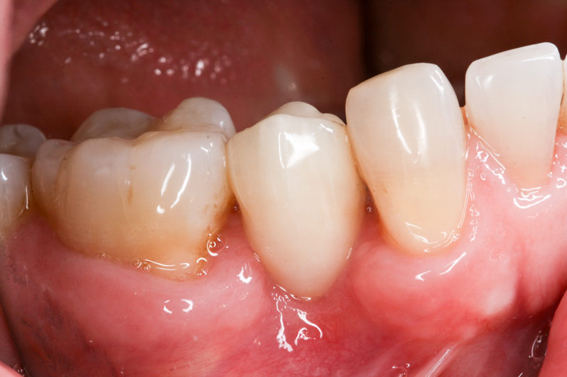
The crown is delivered in two weeks and cemented in place.

This picture was taken 3 months after the crown was cemented, demonstrating the tissue-friendliness of this material despite the deep position of the implant.